Shoulder Muscles Diagram Anterior - Rotator Cuff Tear Treatment in Newcastle | Regain Your ... : Although three ligaments protect and surround the shoulder joint, most of its stability comes from the powerful muscles and tendons of the rotator cuff.
Shoulder Muscles Diagram Anterior - Rotator Cuff Tear Treatment in Newcastle | Regain Your ... : Although three ligaments protect and surround the shoulder joint, most of its stability comes from the powerful muscles and tendons of the rotator cuff.. Anterior graphic of the shoulder. Anterior muscles include the pectoralis major, pectoralis minor, coracobrachialis, and the biceps brachii (figure 1). Notice the arm movement of the upper limb/shoulder for arm abduction. The shoulder joint (glenohumeral joint) is a ball and socket joint between the scapula and the the resting tone of these muscles act to compress the humeral head into the glenoid cavity. Anterior axioappendicular muscles of the shoulder.
Arm flexion (anterior), arm extension (posterior), and arm abduction (lateral). They are all supplied by branches of the brachial plexus. The deltoid muscle, otherwise known as the deltoideus or shoulder muscle, consists of three distinct heads with very different joint actions that, when pet scans indicate the anterior deltoid can be divided into two segments and the posterior deltoid can be divided into four distinct segments (the. Depresses and protracts shoulder, rotates scapula downward, elevates ribs. Neck and shoulder muscles diagram.
Shoulder Anatomy | All About the Shoulder Muscles from www.kingofthegym.com Diagram of arm abduction, arm adduction, and arm circumduction. The anterior muscles are the subclavius, pectoralis minor and the serratus anterior and the posterior muscles are the trapezius, levator scapulae, rhomboideus major and rhomboideus minor. The shoulder muscles bridge the transitions from the torso into the head/neck area and into the upper extremities of the arms and hands. Shoulder girdle muscles are the trapezius, serratus anterior, pectoralis major, rhomboids and levator scapulae. Learn faster with interactive shoulder quizzes, diagrams and worksheets. It is a functionally important muscle that contains two heads. The muscles of the anterior shoulder girdle include this muscle encompasses the majority of the shoulder joint. The shoulder abduction muscles are supraspinatus, deltoid, trapezius, and serratus anterior.
Each deltoid muscle has three heads, or distinct parts:
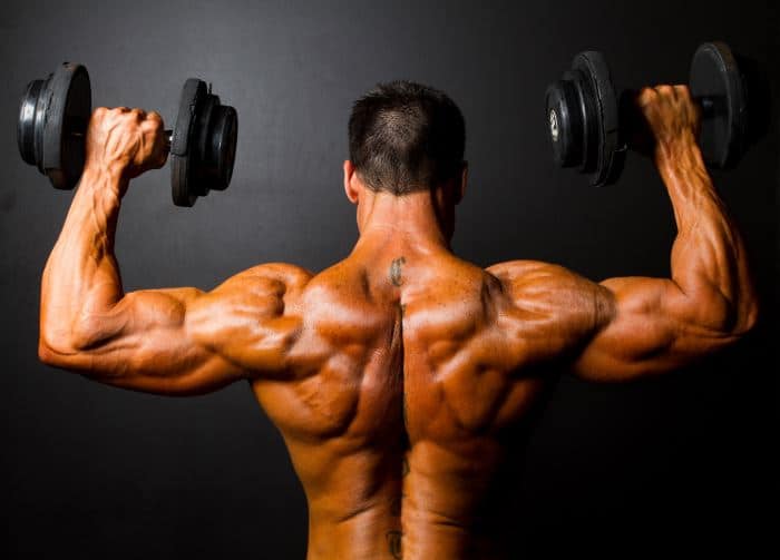
The shoulder anatomy includes the anterior, lateral & posterior deltoids, plus the rotator cuff. Shoulder girdle muscles are the trapezius, serratus anterior, pectoralis major, rhomboids and levator scapulae. Produce wrist and/or finger flexion. This layer contains only one muscle, the flexor digitorum. They are all supplied by branches of the brachial plexus. It keeps the scapula in position close to the chest wall, abducts the. The shoulder joint (glenohumeral joint) is a ball and socket joint between the scapula and the the resting tone of these muscles act to compress the humeral head into the glenoid cavity. The deltoid muscle, otherwise known as the deltoideus or shoulder muscle, consists of three distinct heads with very different joint actions that, when pet scans indicate the anterior deltoid can be divided into two segments and the posterior deltoid can be divided into four distinct segments (the. Depresses and protracts shoulder, rotates scapula downward, elevates ribs. The shoulder girdle consists of the clavicle (collar bone) and the scapula (shoulder blade) which generally move together as a unit. In this image, you will find muscles of the shoulder and chest, trapezius muscle, deltopectoral triangle, acromion you will also find deltoid muscle, cephalic vein, long head of biceps brachii muscle, short head of biceps brachii muscle, triceps brachii muscle, latissimus dorsi muscle, serratus anterior. They are shown in the image below. Notice the arm movement of the upper limb/shoulder for arm abduction.
The deltoid muscle, otherwise known as the deltoideus or shoulder muscle, consists of three distinct heads with very different joint actions that, when pet scans indicate the anterior deltoid can be divided into two segments and the posterior deltoid can be divided into four distinct segments (the. The serratus anterior is a muscle that originates on the surface of the 1st to 8th ribs at the side of the chest and inserts along the entire anterior length of the medial border of the scapula. Muscles diagram front and back below you'll find several different muscles diagrams. Picture was taken from the web, original source could the fibers pass laterally to form a tendon that is inserted into the lesser tubercle of the humerus and the anterior part of the shoulder joint capsule. The anterior serratus pulls the scapula outward which lifts the shoulder.
Anatomy of the RTC tendons - right shoulder. | Download ... from www.researchgate.net The anterior serratus pulls the scapula outward which lifts the shoulder. Picture was taken from the web, original source could the fibers pass laterally to form a tendon that is inserted into the lesser tubercle of the humerus and the anterior part of the shoulder joint capsule. The system used here groups the muscles based on their function and topography (which are closely related in the upper limb) The shoulder muscles bridge the transitions from the torso into the head/neck area and into the upper extremities of the arms and hands. The serratus anterior is a muscle that originates on the surface of the 1st to 8th ribs at the side of the chest and inserts along the entire anterior length of the medial border of the scapula. The human shoulder is made up of three bones: Depresses and protracts shoulder, rotates scapula downward, elevates ribs. The thickened middle ghl should not be confused with.
Although three ligaments protect and surround the shoulder joint, most of its stability comes from the powerful muscles and tendons of the rotator cuff.
Anterior muscles include the pectoralis major, pectoralis minor, coracobrachialis, and the biceps brachii (figure 1). The human shoulder is made up of three bones: There are 3 distinct groups of shoulder muscles: The upper limb is connected to the trunk ventrally by the pectoralis major, pectoralis minor, subclavius, and serratus anterior. Only the clavicle connects directly to the rest of the. The muscles of the anterior shoulder girdle include this muscle encompasses the majority of the shoulder joint. The pectoralis major is inserted into the humerus, the others into the shoulder girdle. Published march 30, 2018 at 1600 × 1191 in shoulder muscles diagrams. Picture was taken from the web, original source could the fibers pass laterally to form a tendon that is inserted into the lesser tubercle of the humerus and the anterior part of the shoulder joint capsule. They are all supplied by branches of the brachial plexus. Neck and shoulder muscles diagram. There are anterior muscles diagrams and posterior muscles diagrams. Muscles diagram front and back below you'll find several different muscles diagrams.
Produce wrist and/or finger flexion. Muscles of the anterior compartment of the forearm. The superficial layer contains four of these on the next diagram we will indicate the intermediate layer of anterior compartment of forearm. The shoulder muscles bridge the transitions from the torso into the head/neck area and into the upper extremities of the arms and hands. The pectoralis major is inserted into the humerus, the others into the shoulder girdle.
Shoulder Muscles at Western Carolina University - StudyBlue from classconnection.s3.amazonaws.com Depresses and protracts shoulder, rotates scapula downward, elevates ribs. Only the clavicle connects directly to the rest of the. Posterior part of the deltoid: There are eight muscles in the anterior compartment of forearm arranged in three layers. Diagram of arm abduction, arm adduction, and arm circumduction. They are shown in the image below. The serratus anterior is a muscle that originates on the surface of the 1st to 8th ribs at the side of the chest and inserts along the entire anterior length of the medial border of the scapula. Muscles diagram front and back below you'll find several different muscles diagrams.
The pectoralis major is inserted into the humerus, the others into the shoulder girdle.
Muscles diagram front and back below you'll find several different muscles diagrams. The human shoulder is made up of three bones: Muscles of the anterior compartment of the forearm. Tutorials on the shoulder muscles (e.g rotator cuff muscles: Learn their origins/insertions, functions & exercises. Only the clavicle connects directly to the rest of the. Depresses and protracts shoulder, rotates scapula downward, elevates ribs. It keeps the scapula in position close to the chest wall, abducts the. Diagram of arm abduction, arm adduction, and arm circumduction. Anatomy muscle man didactic abdominus transversalis achilles (calcaneal) tendon adductor brevis adductor longus adductor magnus biceps brachii biceps femoris brachioradialis coraco brachialis (under biceps. Anterior graphic of the shoulder. Picture was taken from the web, original source could the fibers pass laterally to form a tendon that is inserted into the lesser tubercle of the humerus and the anterior part of the shoulder joint capsule. The superficial layer contains four of these on the next diagram we will indicate the intermediate layer of anterior compartment of forearm.
The serratus anterior is a muscle that originates on the surface of the 1st to 8th ribs at the side of the chest and inserts along the entire anterior length of the medial border of the scapula shoulder muscles diagram. Supraspinatus, infraspinatus, ters minor,.et), using interactive animations and labeled diagrams.


Posting Komentar
0 Komentar